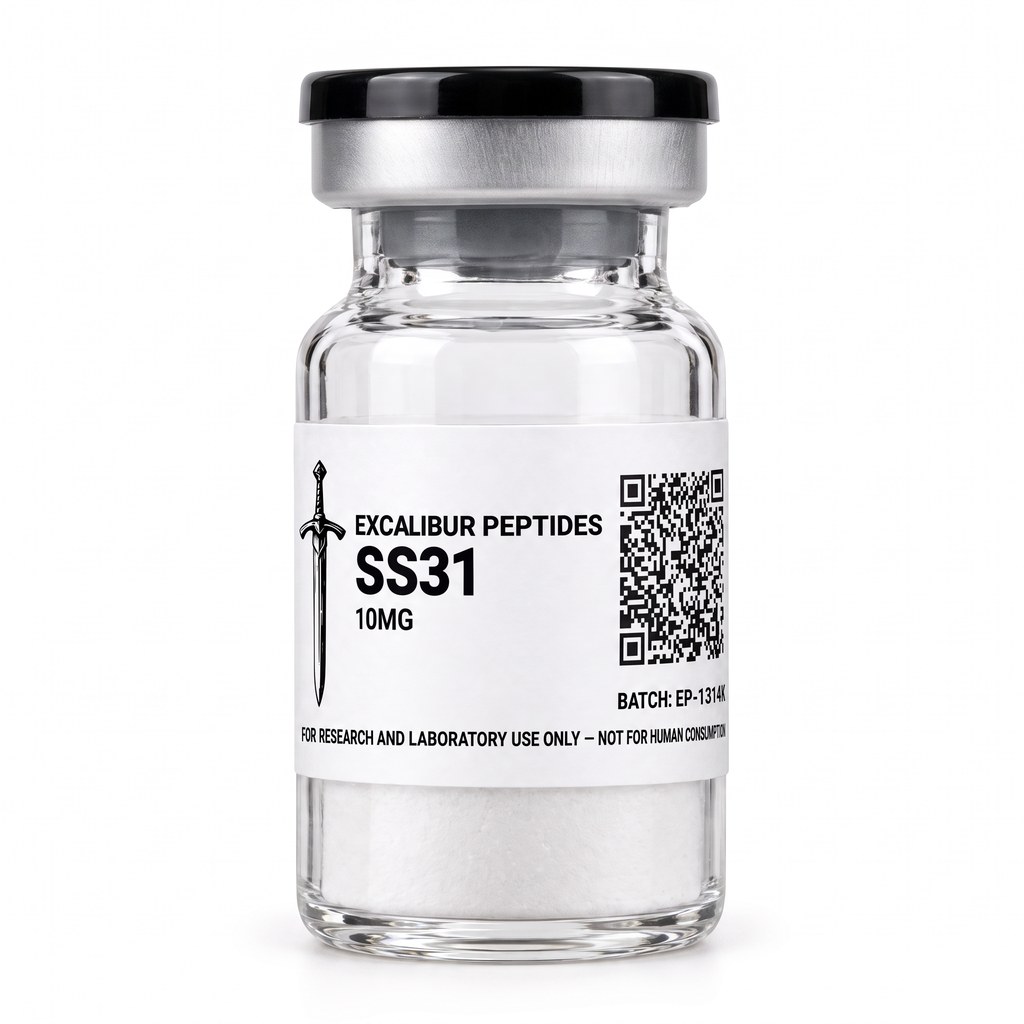
Mitochondrial research spent decades chasing a simple idea: if oxidative damage drives cellular decline, then flooding the cell with antioxidants should slow that decline. Trial after trial in cellular and animal systems failed to reproduce the effect at the level anyone expected. The problem, as it turned out, was not the hypothesis so much as the delivery. Conventional antioxidants distribute throughout the cytosol at concentrations that never approach what is needed inside the organelle where the reactive species are actually generated. SS-31 emerged from a different design philosophy — instead of scavenging radicals wherever they happen to be, target the structure that produces them.
For in-vitro laboratory research use only. Not for human consumption.
What SS-31 Is
SS-31 is a synthetic tetrapeptide belonging to the Szeto-Schiller (SS) class of aromatic-cationic peptides, named for the two researchers whose laboratories characterized the series. The molecule is small — four residues, a molecular weight near 640 g/mol — and carries a distinctive alternating pattern of aromatic and basic residues that gives the class its name and its behavior.
The sequence is D-Arg-Dmt-Lys-Phe-NH₂, where Dmt is 2',6'-dimethyltyrosine. Three structural features matter for how the peptide behaves in experimental systems:
- The D-arginine at position one. Using the D-enantiomer rather than the natural L-form makes the peptide resistant to aminopeptidase cleavage, which is the primary route of degradation for short linear peptides in biological matrices.
- The alternating aromatic-cationic motif. The peptide carries a net charge of +3 at physiological pH, distributed across residues that also present bulky aromatic faces. This combination is what allows it to partition into a phospholipid bilayer while remaining water-soluble.
- The C-terminal amide. Capping the carboxyl terminus removes a negative charge and blocks carboxypeptidase activity, further extending the peptide's half-life in solution.
In the published literature SS-31 also appears under the names elamipretide, MTP-131, and Bendavia, depending on which development program the paper is describing. Researchers searching the literature should query all four terms, because the naming shifted over time and the same molecule appears under different labels in papers separated by only a few years.
The Defining Property: Membrane-Potential-Independent Uptake
Most mitochondria-targeted compounds work by exploiting the electrochemical gradient across the inner mitochondrial membrane. Triphenylphosphonium-conjugated molecules, the best-known example, accumulate inside energized mitochondria because the matrix is negatively charged relative to the cytosol and the lipophilic cation is pulled across. This is elegant, but it has a serious limitation for research on dysfunctional mitochondria: as membrane potential collapses, the compound stops accumulating. The organelles that most need the intervention are precisely the ones that stop taking it up.
SS-31 does not depend on membrane potential. Studies using isolated mitochondria treated with uncouplers such as FCCP — which collapse the proton gradient entirely — still show peptide accumulation in the inner membrane. The mechanism appears to be direct physicochemical partitioning: the aromatic residues insert into the hydrophobic core of the bilayer while the cationic residues remain associated with the anionic phospholipid headgroups at the interface.
This property is the single most important thing to understand about SS-31 in an experimental context. It means the peptide can be studied in models of severe mitochondrial dysfunction where potential-dependent probes fail, and it means concentration inside the organelle is not a simple function of respiratory state.
Cardiolipin: The Molecular Target
The inner mitochondrial membrane contains a phospholipid found essentially nowhere else in the eukaryotic cell: cardiolipin. Structurally, cardiolipin is a dimeric phospholipid — two phosphatidyl moieties linked by a glycerol bridge, producing a molecule with four acyl chains and two negative charges on a conical rather than cylindrical geometry.
That conical shape is functionally important. Lipids with this geometry favor regions of high negative curvature, which is exactly the topology of the cristae — the deep invaginations of the inner membrane where the respiratory complexes are concentrated. Cardiolipin is enriched at cristae, and a substantial body of structural biology shows that it is not a passive bystander. Cardiolipin molecules are resolved in the crystal structures of respiratory complexes III and IV, occupying defined binding pockets. It is required for the proper assembly of respiratory supercomplexes. And it anchors cytochrome c to the inner membrane surface, holding this soluble electron carrier in position between complex III and complex IV.
SS-31 associates selectively with cardiolipin. The interaction has been characterized by NMR, by isothermal titration calorimetry, and by studies in model membranes with defined lipid compositions. The peptide binds cardiolipin-containing bilayers with markedly higher affinity than bilayers composed of neutral phospholipids, and the binding does not simply neutralize charge — it changes the packing behavior of the lipid.
The Cytochrome c Peroxidase Problem
The most mechanistically detailed line of SS-31 research concerns cytochrome c. Under normal conditions, cytochrome c functions as a single-electron shuttle: it accepts an electron from complex III, diffuses along the membrane surface, and delivers it to complex IV. Its heme iron is coordinated by a methionine residue in a compact, closed conformation.
When cytochrome c binds cardiolipin in a particular orientation, that methionine coordination can be disrupted. The heme iron becomes partially exposed, and the protein acquires a secondary activity — peroxidase activity. Instead of transferring single electrons cleanly, it begins catalyzing peroxidation reactions on nearby lipids, including the cardiolipin it is bound to. The peroxidized cardiolipin loses its ability to anchor cytochrome c, releasing it from the membrane. This is a self-amplifying cycle and it appears in the literature as an early event in mitochondrial dysfunction cascades.
Birk and colleagues (2013) demonstrated that SS-31 interferes with this cycle. In their experimental systems the peptide preserved the closed conformation of membrane-bound cytochrome c, suppressed its peroxidase activity, and maintained the cytochrome c-cardiolipin association in a state consistent with normal electron transfer. The proposed mechanism is that SS-31, by occupying cardiolipin interaction sites, shields the specific lipid-protein contacts that trigger the conformational change.
This is a genuinely different mode of action from radical scavenging. SS-31 does not have to be present in stoichiometric excess over reactive oxygen species. It acts upstream, on the structural conditions that permit their generation.
Cristae Architecture and Supercomplex Assembly
A second research domain concerns the physical organization of the inner membrane. Electron microscopy and electron tomography studies in models of mitochondrial stress consistently show cristae disorganization — loss of the tight lamellar stacking, widening of the intracristal space, fragmentation of the membrane network.
SS-31 exposure has been reported to preserve cristae morphology in several such models. The interpretation offered in the literature connects back to cardiolipin: because cardiolipin's conical geometry stabilizes negative curvature, and because SS-31 binding alters cardiolipin packing, the peptide may be acting as a structural stabilizer of the curvature that defines cristae topology.
Downstream of architecture is supercomplex assembly. Respiratory complexes I, III, and IV are not randomly dispersed; they organize into higher-order assemblies — respirasomes — that are thought to improve electron transfer efficiency by reducing the diffusion distance between components. Cardiolipin is required for supercomplex formation. Studies examining SS-31 in models of supercomplex disassembly have reported preservation of the assembled state, measured by blue-native PAGE and by activity-based assays.
The functional consequence measured in these systems is coupling efficiency — the ratio of ATP produced per oxygen consumed. Improved coupling means fewer electrons leak prematurely to molecular oxygen, which means less superoxide generated as a byproduct. This is the throughline that connects the structural observations to the redox observations: SS-31 does not appear to be an antioxidant in the conventional sense, but by maintaining the integrity of the electron transport machinery it reduces the rate at which oxidants are produced in the first place.
Preclinical Research Domains
Skeletal Muscle and Aging Models
Aged skeletal muscle shows characteristic mitochondrial changes: reduced ATP production capacity, lower maximal oxygen consumption rates, and altered redox state. Siegel and colleagues (2013) reported that SS-31 exposure improved mitochondrial energetics in aged mouse skeletal muscle, with measured increases in ATP production and reductions in mitochondrial hydrogen peroxide emission. The magnitude of the effect and the speed of onset in these experiments have made muscle bioenergetics a durable model system for the compound.
Cardiac Tissue Models
Cardiac muscle is among the most mitochondria-dense tissues, with mitochondria occupying roughly a third of the cardiomyocyte volume. Ischemia-reperfusion models are the standard experimental paradigm, and SS-31 has been examined extensively in them. Reported observations include preservation of mitochondrial ultrastructure, maintained ATP levels during reperfusion, and reduced infarct size in the animal models used. The mechanistic reading follows the cardiolipin story — reperfusion is precisely the condition that generates the burst of oxidants that trigger cytochrome c peroxidase activity.
Renal Models
The proximal tubule of the kidney is another high-mitochondrial-density tissue, and models of acute kidney injury have been used to study SS-31. Published work reports preservation of tubular mitochondrial structure and function in ischemic and toxin-induced injury models.
Neuronal and Retinal Models
Neurons cannot store significant energy reserves and depend on continuous oxidative phosphorylation. Models of neurodegeneration frequently show early mitochondrial abnormalities, and SS-31 has been studied in several. Retinal models are of particular interest because the photoreceptor inner segment is densely packed with mitochondria and the tissue is optically accessible for structural imaging.
Primary Mitochondrial Disease Models
Cell lines derived from patients with defined mitochondrial genetic defects provide a well-controlled system for studying whether SS-31 can improve function in mitochondria with a fixed underlying lesion. Results across these systems vary by the specific defect, which is itself informative — it suggests the peptide acts on membrane organization rather than compensating for a missing catalytic function.
Distinguishing SS-31 From Conventional Antioxidants
Researchers designing experiments with SS-31 should be clear about what distinguishes it from the antioxidant compounds it is sometimes grouped with:
| Property |
Conventional antioxidant |
SS-31 |
| Site of action |
Distributed throughout cell |
Concentrated in inner mitochondrial membrane |
| Uptake mechanism |
Passive diffusion / transport |
Direct partitioning, potential-independent |
| Primary activity |
Stoichiometric radical scavenging |
Cardiolipin association, structural stabilization |
| Effect on ROS |
Removes ROS after formation |
Reduces rate of ROS formation |
| Dependence on membrane potential |
N/A |
None |
The practical implication for experimental design is that appropriate controls differ. Comparing SS-31 to a general antioxidant such as N-acetylcysteine tests a different hypothesis than comparing it to another mitochondria-targeted compound, and comparing it to a cardiolipin-binding molecule without the aromatic-cationic motif tests something different again.
Analytical Verification: What a COA Should Show
SS-31 presents specific analytical challenges that a researcher should understand when evaluating a certificate of analysis.
Purity by reverse-phase HPLC. The research-grade floor is ≥99%. For a short peptide like SS-31, the impurity profile is dominated by deletion sequences (a residue missing from the chain) and incomplete deprotection products. Both elute close to the target peak, so the chromatogram should show adequate resolution — a single dominant peak with a clean baseline return, not a broad envelope with unresolved shoulders.
Identity by mass spectrometry. The theoretical monoisotopic mass should be reported alongside the observed mass. This matters more than usual for SS-31 because of the D-arginine and the dimethyltyrosine: a synthesis error that substitutes L-arginine for D-arginine, or tyrosine for 2',6'-dimethyltyrosine, produces a molecule with a different mass. In the L/D case the mass is identical and only chiral analysis would detect it, which is why sourcing from a manufacturer with documented amino acid provenance matters.
Batch specificity. The COA must reference the lot number on the vial. A generic COA showing a representative batch tells you nothing about the material in front of you.
Water content and net peptide content. Lyophilized peptides retain residual water and counterion (typically trifluoroacetate or acetate). The gross vial weight is not the peptide weight. Research requiring accurate molar concentrations should account for net peptide content.
Handling and Storage
Lyophilized SS-31 should be stored at -20°C, protected from light and moisture. The lyophilized form is substantially more stable than any solution, and the material should be kept in that state until it is needed.
General laboratory handling considerations for aromatic-cationic peptides of this class:
- Allow vials to equilibrate to room temperature before opening. Cold glass condenses atmospheric moisture, and water is the primary enemy of a lyophilized peptide's shelf stability.
- Minimize freeze-thaw cycling of any prepared solution. Each cycle promotes aggregation and hydrolysis.
- Use appropriately inert labware. Cationic peptides adsorb to some plastic surfaces, which can silently reduce effective concentration in dilute preparations.
- Protect from light. The dimethyltyrosine residue is a photosensitive aromatic system.
This site does not publish reconstitution protocols, concentration guides, or administration procedures. Those decisions belong to the researcher operating under an approved protocol in a properly equipped facility.
Experimental Readouts Used in SS-31 Studies
Because SS-31 acts on membrane organization rather than on a single enzyme, no one assay captures its effect. The published literature converges on a set of complementary readouts, and researchers designing new work generally combine several of them.
High-resolution respirometry. Oxygen consumption measured in permeabilized fibers or isolated mitochondria, with sequential substrate-uncoupler-inhibitor titration, gives state 3 (ADP-stimulated) and state 4 (leak) respiration rates. The respiratory control ratio derived from these is the classic index of coupling. SS-31 studies commonly report changes in leak respiration rather than in maximal capacity, which is consistent with a mechanism that reduces electron slip rather than adding catalytic capacity.
ATP production rate. Measured either luminometrically or by ³¹P magnetic resonance spectroscopy in intact tissue. Pairing ATP production with oxygen consumption yields the P/O ratio, which is the most direct measure of coupling efficiency and the readout most frequently cited in SS-31 papers.
Mitochondrial hydrogen peroxide emission. Amplex Red-based fluorimetry on isolated mitochondria quantifies the rate of H₂O₂ release. This is the measurement that distinguishes "produces fewer oxidants" from "removes oxidants after production" — a scavenger added to the assay buffer would suppress the signal without changing the underlying generation rate, whereas a structural agent reduces emission at the source. Careful SS-31 experiments include controls that separate these possibilities.
Blue-native PAGE for supercomplex assembly. Solubilizing mitochondrial membranes with digitonin under gentle conditions preserves higher-order respiratory assemblies, which then resolve as discrete bands. In-gel activity staining confirms that the assemblies are catalytically competent, not just physically associated.
Electron microscopy and tomography. Cristae density, intracristal space width, and membrane continuity are measured directly. Tomography is preferred over conventional thin-section EM because the three-dimensional reconstruction avoids sectioning artifacts that can mimic cristae disorganization.
Cardiolipin quantification and oxidation state. Mass spectrometry-based lipidomics quantifies total cardiolipin, its acyl chain composition, and the abundance of oxidized species. Since peroxidized cardiolipin is the proposed intermediate in the cytochrome c cascade, this readout tests the mechanistic hypothesis directly rather than inferring it from downstream effects.
Cytochrome c conformational state. Absorbance spectroscopy in the Soret region, or peroxidase activity assays using a chromogenic substrate, report on whether the heme iron is in the closed methionine-coordinated state or the open peroxidase-competent state.
Common Pitfalls in SS-31 Experimental Design
Several recurring problems appear in work with this compound, and most of them trace back to treating it as if it were a conventional small-molecule antioxidant.
Assuming concentration in the buffer equals concentration in the membrane. SS-31 partitions into cardiolipin-containing bilayers, so its effective local concentration in the inner membrane can be orders of magnitude above the nominal concentration in the medium. Reporting only the buffer concentration makes cross-study comparison difficult and can produce apparently contradictory dose-response relationships between systems with different mitochondrial content.
Using isolated mitochondria without accounting for cardiolipin loss during preparation. Standard differential centrifugation protocols can strip loosely associated lipids and damage outer membranes. Preparations with degraded cardiolipin will respond differently from intact organelles, and preparation quality should be verified before drawing conclusions about the peptide.
Inappropriate comparator compounds. Comparing SS-31 to a cytosolic antioxidant tests the delivery hypothesis, not the mechanism hypothesis. A well-designed study includes at least one other mitochondria-targeted agent so that "targeted delivery" and "cardiolipin-specific action" can be separated.
Ignoring the aromatic residue's photosensitivity. Dimethyltyrosine absorbs in the near-UV. Prolonged exposure of stock solutions to laboratory lighting, particularly in clear tubes, degrades the peptide. Amber vials and minimized bench time are standard practice.
Freeze-thaw cycling. Aromatic-cationic peptides aggregate readily. An aliquoting strategy set up at the time of first reconstitution avoids the silent concentration losses that accumulate over repeated cycles and that show up in the data as increasing variance across a study's timeline.
Adsorption losses in dilute preparations. At low nanomolar working concentrations, a meaningful fraction of a +3-charged peptide can adsorb to standard polypropylene surfaces. Low-binding labware, or carrier protein where the experiment tolerates it, prevents an effect that otherwise looks like inconsistent potency.
Reading the SS-31 Literature Critically
The SS-31 literature spans roughly two decades and includes work from cell-free systems, isolated organelles, cultured cells, multiple animal models, and human clinical programs. A few habits help in evaluating it.
Check whether the system had a cardiolipin defect. The proposed mechanism predicts that effects should be largest where cardiolipin-dependent processes are impaired and smallest in healthy, well-coupled mitochondria. Studies reporting no effect in a healthy control system are not contradictory results — they are consistent with the mechanism.
Distinguish acute exposure from chronic exposure. Structural stabilization effects appear rapidly in isolated systems; changes in mitochondrial biogenesis or content require longer timescales and involve different signaling. Papers reporting increased mitochondrial mass after prolonged treatment are describing an adaptive response, not the primary molecular action.
Watch for the naming problem. Because the same compound appears as SS-31, elamipretide, MTP-131, and Bendavia, a literature search on a single term returns a biased slice of the field, and the slice tends to correlate with the era and the sponsor. Meta-analyses that miss this produce incomplete pictures.
Separate structural endpoints from functional endpoints. Preserved cristae morphology and improved P/O ratio are related but not identical claims. Papers that measure only one and infer the other are making a reasonable but unverified inference.
Note the model species and tissue. Mitochondrial composition, cardiolipin acyl chain profile, and supercomplex organization vary between species and between tissues within a species. An effect size measured in mouse cardiac mitochondria does not transfer directly to a human fibroblast line.
Where SS-31 Sits in the Research Landscape
The compound occupies a specific niche. It is not a broad metabolic modulator like AMPK activators, and it is not a redox buffer like glutathione precursors. It is a structural agent that acts on one phospholipid in one membrane. That narrowness is a feature for experimental design — it makes the mechanistic hypothesis testable in a way that broader-acting compounds do not permit.
For researchers comparing mitochondrial research tools, the useful contrast set includes MOTS-c (a mitochondrial-derived peptide that acts through AMPK and retrograde nuclear signaling rather than membrane structure) and NAD+ (a redox coenzyme and sirtuin substrate acting on metabolic flux). All three are studied in mitochondrial contexts, but they engage completely different mechanisms, and using them as if they were interchangeable produces uninterpretable data.
Why Synthesis Quality Matters More Than Usual Here
Most research peptides are built entirely from standard L-amino acids using well-optimized solid-phase Fmoc chemistry. SS-31 is not. Two of its four residues are non-standard, and each introduces a specific failure mode that a careless synthesis will produce and a superficial certificate of analysis will not catch.
D-arginine. Incorporating the D-enantiomer requires purchasing the D-form building block, which costs several times more than the L-form. A substitution — deliberate or accidental — produces a peptide with an identical molecular formula, identical mass, and nearly identical HPLC retention behavior under standard achiral conditions. Only chiral chromatography or amino acid analysis after hydrolysis will detect it. The functional consequence is significant: the L-form is a substrate for aminopeptidases, so the material degrades far faster in any biological matrix, and experiments that depend on stability over hours will produce results that do not reproduce.
2',6'-dimethyltyrosine. Dmt is a specialty residue and is meaningfully more expensive again. Substituting plain tyrosine produces a molecule 28 Da lighter, which mass spectrometry will detect immediately — provided the MS data is actually acquired on the shipped lot and the theoretical mass is reported alongside the observed. A certificate that reports only "identity confirmed" without the numbers gives the researcher nothing to check.
Coupling difficulty at the sterically hindered positions. The dimethylation on the tyrosine ring adds steric bulk that slows the coupling reaction at that position. Under compressed synthesis schedules, incomplete coupling produces deletion sequences — chains missing the Dmt residue entirely. These are shorter, lighter, and typically elute earlier on reverse-phase HPLC, but if the gradient is too steep they co-elute with the target and inflate the apparent purity number.
C-terminal amidation. The peptide terminates in an amide, not a free acid. Using the wrong resin, or incomplete cleavage chemistry, yields the free-acid form: one Dalton heavier, one additional negative charge, and a different interaction profile with the anionic cardiolipin headgroup that the whole mechanism depends on.
The practical conclusion is that a purity percentage in isolation is not sufficient documentation for this compound. The chromatogram shape, the numeric mass comparison, and the lot match all carry information that the summary number does not. A supplier that publishes the full analytical package is making it possible for a researcher to catch these problems. A supplier that publishes a percentage is asking to be trusted.
Sourcing Considerations
The quality problems that affect the research peptide market apply with particular force to SS-31, for a specific reason: the two non-standard residues. D-arginine and 2',6'-dimethyltyrosine are more expensive starting materials than standard L-amino acids, and both require careful coupling conditions. A supplier cutting cost on raw materials or on synthesis time will produce material that fails on chirality or on the dimethylation, and neither failure is visible in a purity number alone.
What to require:
- Independent third-party testing, not in-house data. A laboratory with no financial interest in the result.
- Both HPLC and MS on the same lot. Purity without identity is meaningless; identity without purity is equally so.
- The actual chromatogram, not just a summary number. The shape of the trace carries information the percentage does not.
- Lot-matched documentation. The number on the COA and the number on the vial must agree.
- Cold-chain handling. Peptides shipped without temperature control in summer heat arrive degraded regardless of how they left the facility.
Excalibur Peptides supplies SS-31 at ≥99% HPLC-verified purity with an independent batch-level certificate of analysis, shipped from a United States facility. Every compound in the catalog is held to the same ≥99% purity floor, and material that does not meet it is not released.
Frequently Asked Questions
What is SS-31?
SS-31 is a synthetic aromatic-cationic tetrapeptide (D-Arg-Dmt-Lys-Phe-NH₂) that concentrates in the inner mitochondrial membrane and associates selectively with cardiolipin. It is studied in preclinical systems for its effects on cristae architecture, electron transport coupling efficiency, and cytochrome c peroxidase activity. It is supplied strictly for in-vitro laboratory research and is not for human consumption.
Is SS-31 the same as elamipretide?
Yes. SS-31, elamipretide, MTP-131, and Bendavia all refer to the same tetrapeptide. The different names reflect different stages and sponsors of the research history. Literature searches should include all four terms.
How does SS-31 differ from other mitochondria-targeted compounds?
Most mitochondria-targeted compounds accumulate by exploiting the membrane potential, so uptake falls as mitochondria become dysfunctional. SS-31 partitions into the inner membrane independently of membrane potential and acts through cardiolipin association rather than radical scavenging.
What purity should research-grade SS-31 meet?
The research-grade floor is ≥99% by reverse-phase HPLC, with identity confirmed by mass spectrometry against the theoretical molecular weight. Both tests should appear on a lot-specific certificate of analysis from an independent laboratory.
How should SS-31 be stored?
Lyophilized SS-31 should be stored at -20°C, protected from light and moisture, and kept in the lyophilized state until required. Repeated freeze-thaw cycling of prepared solutions should be avoided.
Is SS-31 approved for human use?
No. SS-31 is sold strictly for in-vitro laboratory research and identification purposes. It is not approved by the FDA for human therapeutic use, is not intended for veterinary administration, and is not intended for any clinical application.
View the SS-31 product page · Browse the research catalog · View the COA database
All compounds referenced in this article are sold for in-vitro laboratory research purposes only. They are not drugs, supplements, or medical products, and are not intended for human use, veterinary use, or consumption of any kind. Always consult applicable regulations regarding peptide research in your jurisdiction.
Last updated: July 2026